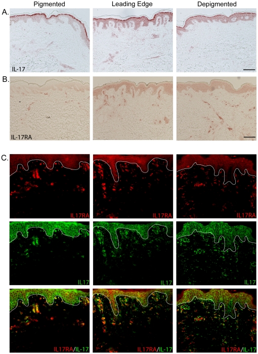
Figure 5. IL-17A and IL-17RA are found on vitiligo skin biopsies.
Immunohistochemistry and immunofluorescence staining of IL-17A and IL-17 receptor A on vitiligo skin biopsies. (A)&(B) In immunohistochemistry, IL-17A and IL-17RA showed strong staining on the upper dermis of leading edge vitiligo skin when compared to non-lesional/pigmented vitiligo skin. (C) Double immunofluorescence staining of IL-17A and IL-17RA, areas of orange shows receptor bound IL-17A molecules. 50–60% of IL-17RA positive cells are also IL-17A positive in leading edge vitiligo biopsies. In comparison, only 20–30% of IL-17RA positive cells are IL-17A positive in non-lesional and lesional skin. Bar = 100 mm, applies to 5A, B&C.

