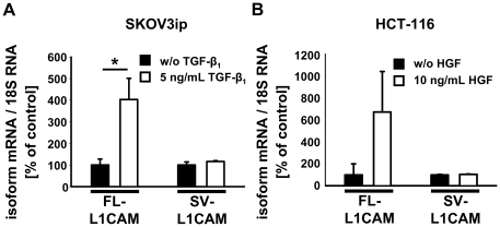
Figure 2. Expression of L1CAM splice variants was deregulated in carcinoma cells upon exposure to pro-metastatic factors.
Mean FL-L1CAM or SV-L1CAM mRNA levels ± SEM (columns ± bars) after incubation with pro-metastatic factors. Target gene mRNA levels were normalized to 18S rRNA levels and the means of the relative reference group without incubation with a pro-metastatic factor were set as 100%. A. 1×10⁵ SKOV3ip-lacZ ovarian carcinoma cells were incubated for 48 h with or without 5 ng/ml of recombinant TGF-β1 (recTGF-β1). FL-L1CAM/Ø: 100.0%±27.6%, n = 9 cell pools; FL-L1CAM/recTGF-β1: 403.5%±98.1%, n = 9 cell pools; SV-L1CAM/Ø: 100.0%±14.2%, n = 9 cell pools; SV-L1CAM/recTGF-β1: 116.2%±4.0%, n = 9 cell pools (FL-L1CAM, SKOV3ip-lacZ+recTGF-β1 vs. SKOV3ip-lacZ−recTGF-β1: **p = 0.005; SV-L1CAM, SKOV3ip-lacZ+recTGF-β1 vs. SKOV3ip-lacZ−recTGF-β1: p = 0.266, as determined by Wilcoxon Signed Rank test). B. 1×10⁵ HCT-116 colon carcinoma cells were incubated for 10 days with or without 10 ng/ml of recombinant HGF (recHGF). FL-L1CAM/Ø: 100.0%±100.0%, n = 9 cell pools; FL-L1CAM/recHGF: 673.9%±369.7%, n = 9 cell pools; SV-L1CAM/Ø: 100.0%±4.6%, n = 9 cell pools; SV-L1CAM/recHGF: 104.9%±2.7%, n = 9 cell pools (FL-L1CAM, HCT-116+recHGF vs. HCT-116−recHGF: p = 0.313; SV-L1CAM, HCT-116+recHGF vs. HCT-116−recHGF: p = 0.250, as determined by Wilcoxon Signed Rank Test).

