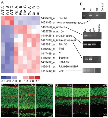
Figure 5. Differential gene expression in P4 retina.
A. Heat map of genes differentially expressed between both mutants and WT selected from the microarray assay. Note that only one gene other than Chrnb2 is downregulated in the mutants (Pisd-ps3). Redundant probesets have been stripped from the map. When a gene symbol is available it appears next to the probeset. Gene names are in Table S4. B. Validation qualitative RT-PCRs for some of the genes in (A). “Control” is without template. Primers used are given in Table 1. Amplification with Crb1 primers yields three bands. The Expasy (www.expasy.org) annotation for mouse Crb1 gives four known splice variants. C. Confocal images of P4 retina using an antibody to CCL21 (aka 6CKine). CCL21 protein is strongly expressed in the outer segment (OS), and outer plexiform layers (OPL) and weakly in the inner plexiform layer (IPL) of Chrnb2−/− but not WT retinas. D. Confocal images of adult retina using an antibody to CCL21 (aka 6CKine). CCL21 protein appears in the outer segment (OS) and outer plexiform layer (OPL) of all animals. Anti-CCL21 (red), DAPI nuclear counterstain (pseudo colored green). Scale 25 µm.

