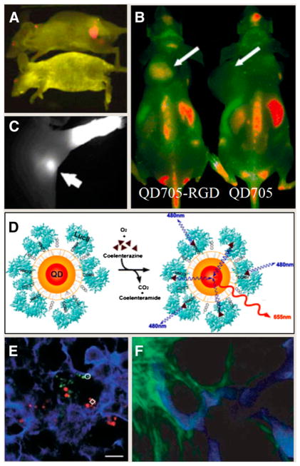
FIGURE 1.
(A) Molecular targeting and in vivo imaging of prostate tumor using QD–antibody conjugate. (Reprinted with permission of (5).) (B) In vivo near-infrared fluorescence imaging of tumor-bearing mice injected with QD705-RGD peptide or QD705. (Reprinted with permission of (7).) (C) NIR QDs taken up by sentinel lymph nodes. (Reprinted with permission of (8).). (D) Principle of self-illuminating QDs through bioluminescence resonance energy transfer of Luc8 acting as donor to acceptor QDs on oxidation of coelenterazine. (Reprinted with permission of (10).) (E) Simultaneous tracking of different populations of QD-labeled metastatic tumor cells in mice lung tissue. (Reprinted with permission of (11).) (F) Intravital imaging provides clear separation of QD-labeled tumor vessels (blue) from green fluorescent protein–expressing perivascular cells (green). (Reprinted with permission of (12).)

