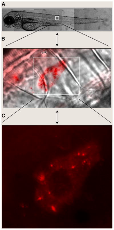
FIGURE 2.

In vivo QD imaging in live zebrafish embryos. Two- to 4-cell–stage embryos were injected with red QDs and were allowed to develop to the larva stage. Live fish were observed in DIC (A and B) or epifluorescence (B and C) on an inverted microscope using a charge-coupled device camera. The brightness and photostability of QDs reveal the dynamic diffusion of single vesicles over a continuum of length scales from whole animals (millimeters) to subcellular levels (less than a micrometer).
