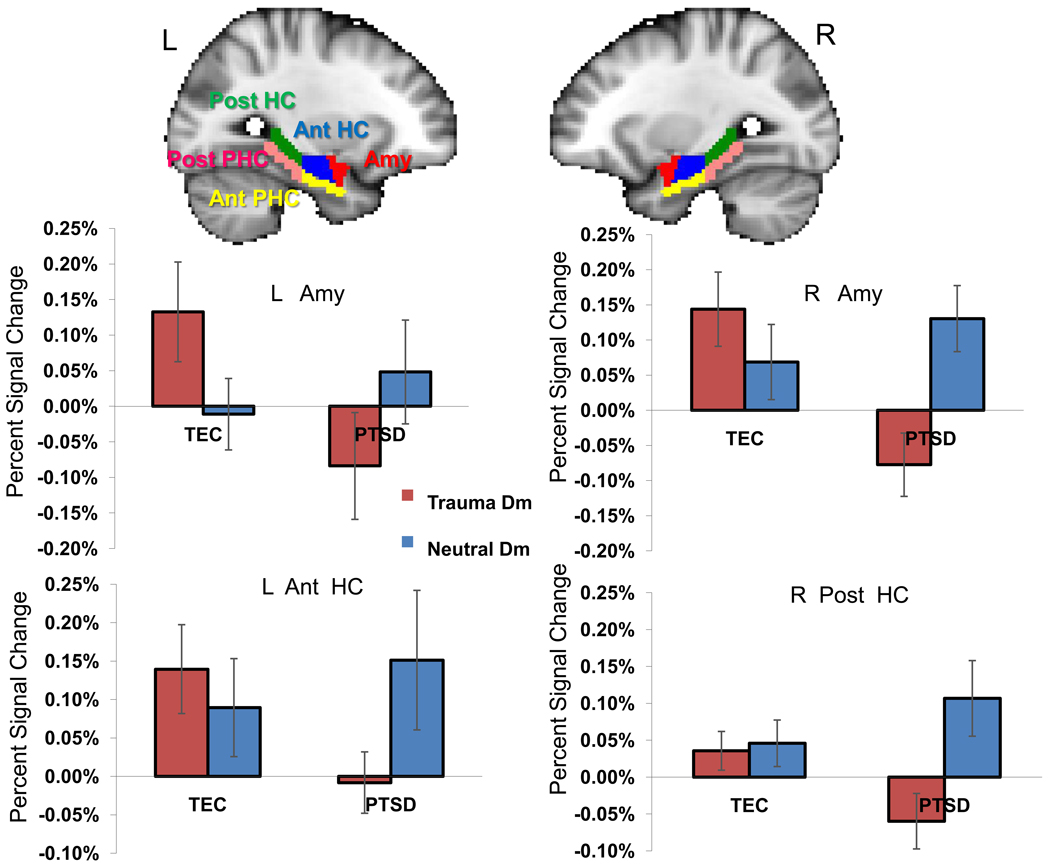
Figure 2.
Region-of-interest analysis results indicate reduced Dm effect for trauma pictures in PTSD group in bilateral amygdala, left anterior hippocampus and right posterior hippocampus. L = left, R = right, Dm = Difference due to memory effect, TEC = Trauma-exposed control group, Amy = amygdala, Ant = Anterior, HC = Hippocampus, PHC = Parahippocampal cortex, Post = Posterior. Error bars represent the standard error of means.

