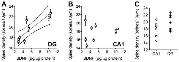
Figure 1. Variability in BDNF protein expression correlates with dendritic spine density in the dentate gyrus but not CA1 subfield of the mouse hippocampus.
(A) BDNF protein levels are positively correlated with dendritic spine density along the secondary and tertiary dendrites of Golgi-impregnated granule cells in the hippocampal dentate gyrus. (B), No such correlation was detected in the CA1 subfield. For both graphs, each symbol represents one mouse, and error bars depict the s.e.m. derived from the variability between cells from each mouse. For the dentate gyrus only, the Pearson’s correlation was significant at p < 0.01. (C), Dendritic spine densities are similar along the apical dendrites of CA1 pyramidal neurons and dentate gyrus granule cells.

