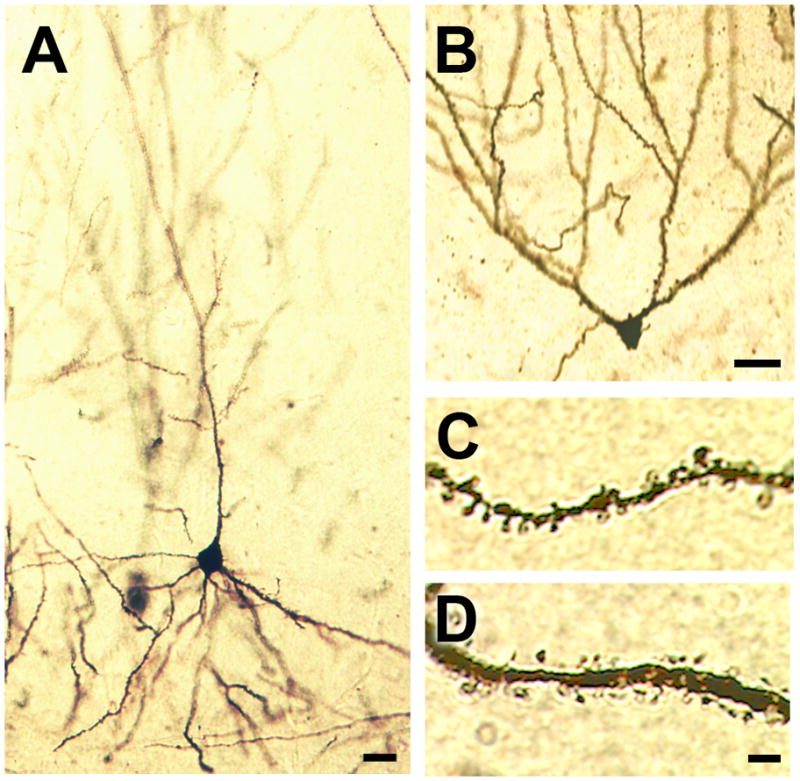
Figure 2. Representative images of golgi-impregnated cells in the mouse hippocampus.

(A), CA1 pyramidal neuron visualized with Golgi impregnation. (B), Dentate gyrus granule cell in the mouse hippocampus. For panels (A–B), scale bar = 20μm. (C), Apical oblique dendritic segment from a CA1 pyramidal neuron in the mouse hippocampus. (D), Secondary dendrite from a Golgi-impregnated dentate gyrus granule cell. For panels (C–D), scale bar = 5.0μm.
