
Figure 2. Concentration-dependent effects of BoNT/A on SNAP-25 protein cleavage in mouse ES-derived motoneurons, primary mouse spinal neurons and DRG neurons, and the motoneuron like NSC-34 cell line.
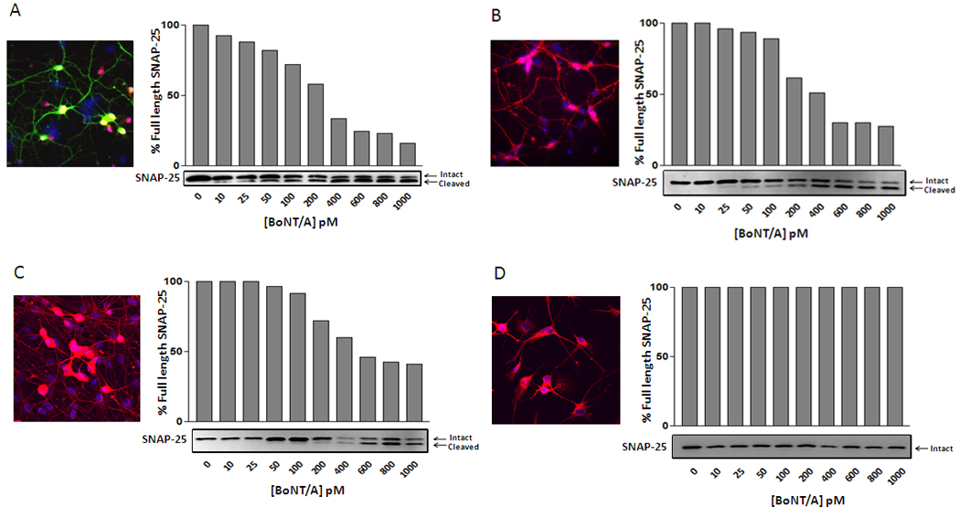
A–D; Graphic representation of the percentage of uncleaved SNAP-25 protein following cell treatment with various concentrations of BoNT/A (0–1000 pM). The graphs represent the quantification of each blot shown at the bottom of the x-axis. The top band is uncleaved SNAP-; the bottom band is the BoNT/A mediated cleavage fragment. Inset in each panel are representative image of : (A) ES-derived motoneuron cultures stained with the neuronal marker NeUN (red), Hb9-eGFP (green) and DAPI (blue); (B) primary mouse spinal cord cells and (C) DRG neurons stained with cytoplasmic neuronal markers Tuj1 (Red) and DAPI (Blue); (D) immortalized NSC-34 cells stained with neuronal marker Tau5 (Red) for total Tau protein and DAPI imaging. The scale bar is 50 µm.