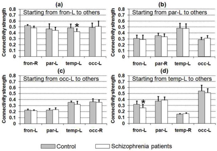
Fig. 3.
CSCP values from frontal (a), parietal (b), occipital (c), and temporal lobes (d) are shown for normal controls (shaded bar) and schizophrenia patients (white bar). Unique CSCP measures from different lobes can be observed. The CSCP is also sensitive to the structural connectivity changes and significantly smaller CSCPs between the temporal lobe and the frontal lobe, highlighted with asterisk in (a) and (d), can be identified in the schizophrenia group.

