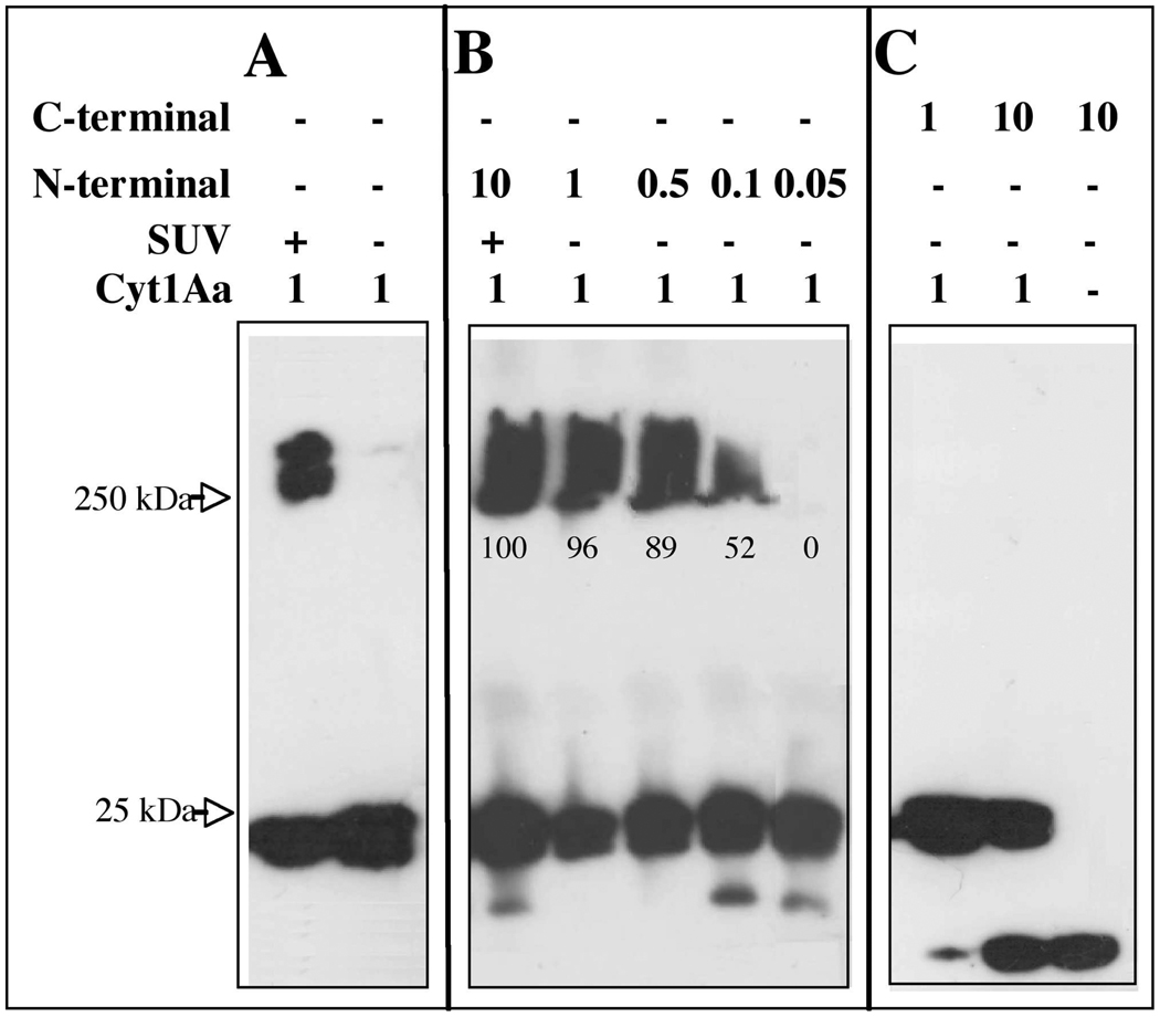
FIGURE 3. Analysis of Cyt1Aa oligomer formation by Western blot using polyclonal anti-Cyt1Aa/11 antibody and a secondary Goat-HRP antibody.
Panel A, Cyt1Aa protoxin was activated with proteinase K in the presence or absence of SUV (PC:Ch:S mixture) liposomes. Panel B, analysis of the effect of N- terminal fragment at different molar ratios on oligomerization of Cyt1Aa in absence of lipid membranes. N-terminal fragment triggers aggregation of Cyt1Aa in absence of lipid membranes. Panel C, analysis of the effect of C- terminal fragment at different molar ratios on oligomerization of Cyt1Aa in absence of lipid membranes. Size of proteins was estimated from molecular pre-stained precision plus standard, all blue (BioRad). Numbers within the images represent the percentage of aggregate that was formed in the presence of N-terminal fragment as determine by scanning optical density of bands in the blots

