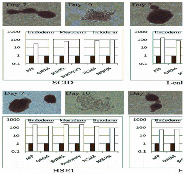
Figure 3. Differentiation of PID-specific iPSCs reveals lineage-specific gene expression.
PID-specific iPSC were allowed to differentiate into embryoid bodies (EB) by culture in a bFGF-free hES medium and without co-culture with feeder cells. Robust formation of tight and well-formed cell clusters was detected by day 7, that became cystic by day 10 (upper row in each cell-specific panel). Quantitative RT-PCR gene expression analysis of the derived EB after 10 days shows increased expression of lineage-specific markers from each of the three embryonic germ layers, including: AFP and GATA4 (endoderm), RUNX1 and Brachyury (mesoderm), NCAM and NESTIN (ectoderm). Quantitative RT-PCR reactions were normalized against β-actin (ACTB). Expression was calculated using the ddCT method relative to expression levels in undifferentiated iPSCs. Black and white bars identify undifferentiated iPSCs and day 10 EB, respectively.

