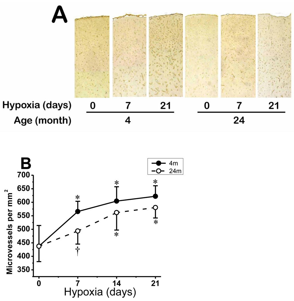
Fig. 7.
Microvascular density in young and aged mice cerebral cortex in normoxic control and prolonged hypoxia. A: composite photomicrograph of GLUT-1–stained sections spanning part of the parietal cortex of 4 and 24 month old mice at normoxia (0), and 7 and 21 days of hypoxia. B: Capillary density count (number per mm2) of GLUT-1-stained sections of 4 month old and 24 month old mice during normoxia (0) and 7, 14 and 21 days of hypoxia. *p < 0.05 compared with corresponding normoxic control. †p < 0.05 compared with corresponding 4 month old mice value. Values are mean ± SD, n = 5 mice per time point in each group.

