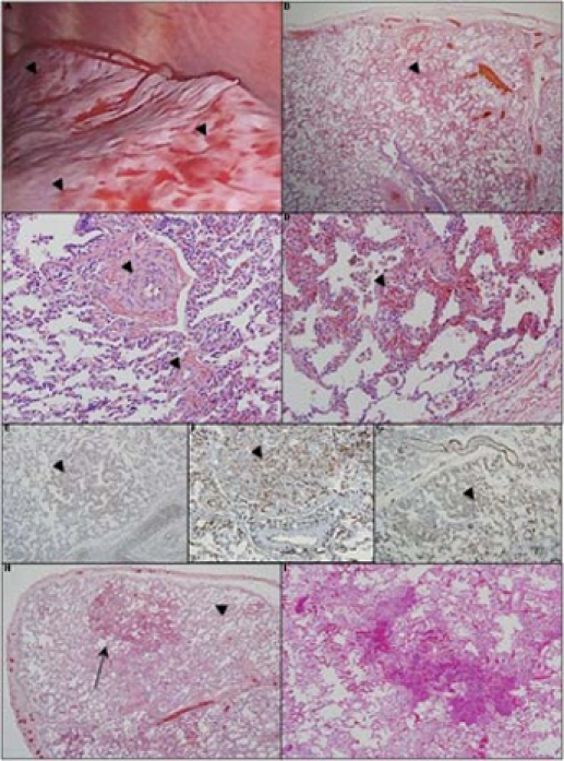
Figure 2.

Intraoperative view and photomicrographs of a lung wedge biopsy section. (A) Intraoperative view shows a grossly nodular appearance (arrowheads). (B) Microscopically there is a patchy distribution of dense cellular interstitial infiltrates, haemorrhagic areas and vascular congestion (arrowhead). (C) The pulmonary vasculature shows muscularisation of small and medium arteries with medial hypertrophy (arrowheads). (D) High power views show vascular congestion with foci of increased capillary density (arrowhead) consistent with capillary haemangiomas. The capillary haemangioma appear to be comprised of endothelial cells without nuclear atypia, which on immunohistochemistry, stain positive (arrowheads) for (E) CD-31, (F) CD-34 and (G) Factor VIII staining. The section did not stain positively for MIB-1 (not shown). Comparison of lung biopsy to autopsy histology shows progression of capillary growth over time. (H) Histology at diagnosis showed a heterogeneous distribution of affected areas (arrow) with sparing of parts of the distal lung parenchyma (arrowhead). (I) Despite interferon α 2a therapy, at the time of death, there was progression in the size of the lesions and a decrease in the unaffected areas of lung. Some of the alveolar infiltrate is due to resuscitative efforts near the time of death.
