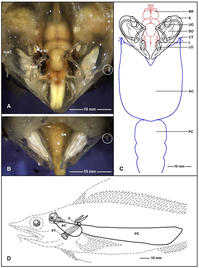
Figure 4.
Inner ears, brain, and swim bladder of Antimora rostrata (anterior to the top for A, B, and C). A: Dorsal view of the brain and inner ears after removing part of the skull and cartilage. B: Ventral view of the inner ears and the brainstem after removing part of the bottom of the cranium, with a clear indication of the rigidity of the saccule without the support of water. After removing the two anterior chambers of the swim bladder from where they attach to the inner ear’s bony capsules, the stubs of the dense connective tissue (indicated by white circles) indicate the attachment points. C: The relationship between the brain, inner ears, and swim bladder in Antimora; note that the size of the brain is relatively small compared with the size of the inner ears. D: Lateral view of the relative position between the inner ear and the swim bladder with respect to the head and the body. Also shown in D are two vertebrae (V) and a muscle bundle to the upper pharyngeal teeth (PT). AC, PC, anterior and posterior chamber of swim bladder; A-SC, H-SC, P-SC, anterior, horizontal, and posterior semicircular canals; BR, brain; L, lagena; LO, lagenar otolith; PT, pharyngeal teeth; S, saccule; SN, nerve to saccule; SO, saccular otolith; CT, connective tissue; U, utricle; UO, utricular otolith.

