
Figure 2.
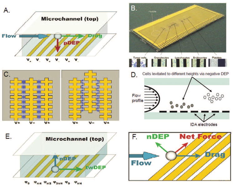
(A) Interdigitated array (IDA) electrodes. (B) Electrosmear slide showing fractionation tumor cells and blood components. Reproduced from Cristofanilli et al. (2008). (C) Castellated IDA electrodes. (D) DEP field-flow fractionation operates by levitating cells against gravity to different heights in the channel via negative DEP, allowing separation to be achieved based on their differing flow velocities. (E) Configuration and forces in a twDEP electrode array. (F) Summation of forces near an angled electrode.