Figure 3.
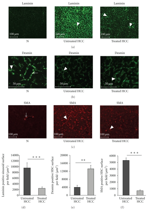
Rosuvastatin prevents sinusoid capillarisation in HCC livers. (a) Representative images of laminin immunostaining in normal livers (N, n = 4), untreated HCC, and rosuvastatin-treated HCC livers (n = 4/group) at 16 weeks. Laminin was expressed by the sinusoids (white arrowhead) only in HCC livers. A weaker expression of laminin was observed in treated-HCC livers compared with untreated HCC. (b) Representative images of desmin immunostaining in normal livers (N, n = 4), in untreated HCC, and in rosuvastatin-treated HCC livers (n = 4/group) at 16 weeks. Desmin was expressed in mural cells lining vessels derived from the portal tract (VDPT) in normal livers (white arrow). Desmin was expressed in nonactivated hepatic stellate cells (HSCs) surrounding the sinusoids in both livers (white arrowhead). (c) Representative images of SMA immunostaining in normal livers (N, n = 4), in untreated HCC, and in rosuvastatin-treated HCC livers (n = 4/group) at 16 weeks. SMA was expressed by smooth muscle cells lining arteries (white arrow). SMA was expressed in activated HSCs surrounding the sinusoids in HCC livers (white arrowhead). (d) Quantification of laminin-positive sinusoid surface per field, in untreated HCC and in rosuvastatin-treated HCC livers (n = 4: group) at 16 weeks. Data are mean ± SEM, ***P < .001. (e) Quantification of desmin-positive hepatic stellate cell (HSC) surface area per field in untreated HCC and in rosuvastatin-treated HCC livers (n = 4/group). Data are mean ± SEM, **P < .01. (f) Quantification of SMA-positive HSC surface area per field in untreated HCC and in rosuvastatin-treated livers (n = 4/group). Data are mean ± SEM, ***P < .001.

