Figure 7.
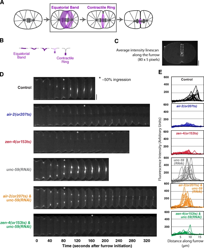
Inhibition of septinUNC-59 rescues the contractile ring assembly defect in Aurora BAIR-2– but not MKLP1ZEN-4-inhibited embryos. (A and B) The schematics highlight the second step in cytokinesis (A, boxed area), assembly of the contractile ring. During this step, the cortex folds in, and contractile ring proteins become concentrated in a compact ring that sits at the furrow tip and are cleared from the remainder of the equatorial cortex (illustrated in B; also see Fig. S3). (C) A schematic of the method used to analyze myosin accumulation at the furrow tip. At the time point when the furrow had closed to ∼50% of its initial diameter, a mean linescan (80 pixels long and 5 pixels wide) was drawn from the edge of the embryo along the furrow toward the tip. (D) Representative montages of the furrow region for each condition are shown. The asterisks mark the time point when the furrow had closed to ∼50% of its initial diameter. (E) The individual linescans for all embryos (after subtraction of cytoplasmic background) are plotted versus distance along the furrow. All imaging was performed using the postmeiotic upshift conditions outlined in Fig. 2 A. Bars, 10 µm.

