Figure 2.
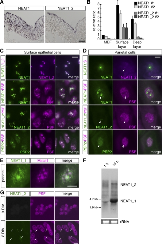
Paraspeckles are formed only in a small subpopulation of cells expressing NEAT1_2. (A) Expression pattern of NEAT1 and NEAT1_2 in the zymogenic region of the adult stomach detected by in situ hybridization. NEAT1_2 expression is restricted to the surface epithelial cells facing the lumen of the stomach. (B) qPCR analysis of the expression of NEAT1 and NEAT1_2 in the dissected surface and deep layer of the gastric epithelium. (C and D) Subnuclear distribution of NEAT1, NEAT1_2 detected by FISH, and the paraspeckle marker PSF and PSP2 in the surface epithelial cells (C) and parietal cells (D). Arrowheads in D indicate the putative transcription sites of NEAT1_2. (E) Different expression of NEAT1_1 and Malat1 in parietal cells that lack expression of NEAT1_2. (F) Induction of NEAT1 expression in cultured MEFs. (G) Paraspeckle formation is rapidly induced in MEFs cultured in vitro (DIV, days in vitro). Bars: (A) 100 µm; (C, D, and G) 10 µm; (E) 5 µm.

