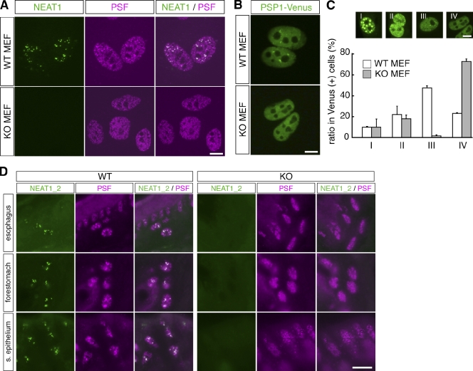
Figure 4.
Paraspeckles are not formed in NEAT1 knockout mice. (A) Subnuclear localization of NEAT1_2 and the paraspeckle marker PSF in MEFs. The foci of PSF were not observable in MEFs from NEAT1 knockout mice. (B) Typical distribution of PSP1-Venus in MEFs from wild-type (WT) and knockout (KO) mice. The paraspeckle marker accumulated as discrete foci in WT MEFs, but not in knockout MEFs. (C) Quantitative analysis of subnuclear localization of PSP1-Venus in WT and knockout MEFs. Overexpressed PSP1-Venus signals were categorized as types I–IV. Note that typical paraspeckle-like distribution (type III) was rarely observed in knockout MEFs. (D) Loss of punctate signals of paraspeckle marker PSF in epithelial cells of esophagus, forestomach, and surface epithelium (s. epithelium) of zymogenic stomach in the knockout mice. Bars, 10 µm.

