Figure 1.

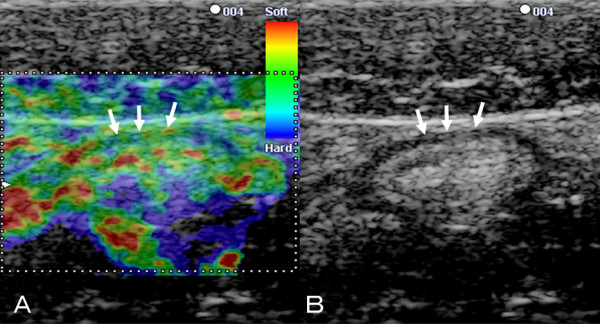
B-mode ultrasound and corresponding EG images of the normal colon wall. A: Corresponding EG of the normal wall shows almost all parts of the wall appear green (arrows)..B: B-mode ultrasound image of the normal colon wall shows 5 layers (arrows).
