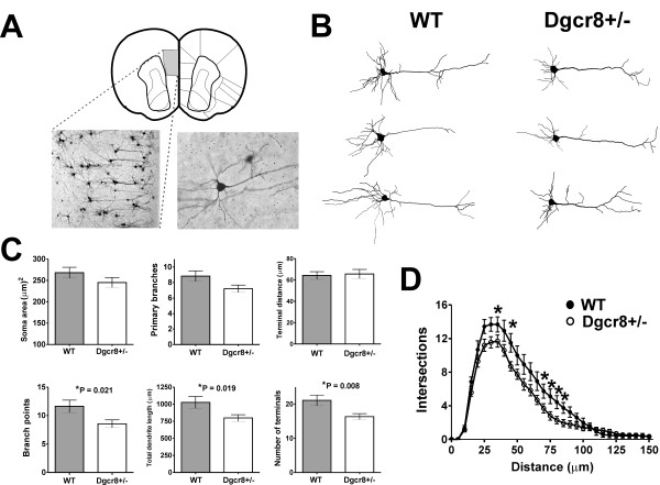
Figure 4.
Reduced basal dendritic complexity of L5 pyramidal neurons in Dgcr8+/- mice. Golgi-cox staining of mPFC from WT and Dgcr8+/- mice at P25. (A) Diagram of coronal section of mouse mPFC delineating the area of study and representative 10× and 40× magnification images of L5 pyramidal neurons. (B) Traces from Neurolucida reconstructions are shown for three representative WT and Dgcr8+/- neurons. (C) Summaries of morphometric data from WT (n = 16 neurons) and Dgcr8+/- mice (n = 20 neurons). Cell body area, number of primary basal dendrites, and the average basal terminal distance from soma were not different between genotypes. Statistically significant decreases were observed in the number of basal dendrite branch points, total dendritic length, and number of terminals. Bars represent mean ± standard error; *P < 0.05. (D) Scholl analysis of basal dendrites shows reduced complexity in Dgcr8+/- neurons; *P < 0.05.

