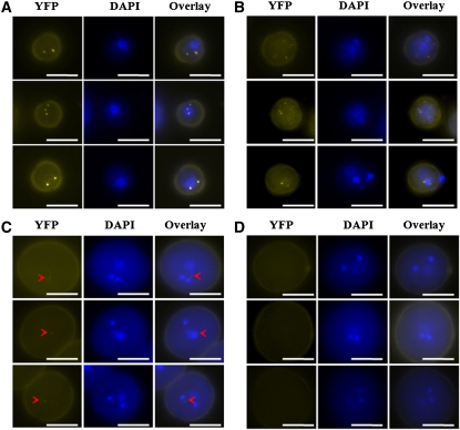
Figure 3.
Subcellular Localization of APC8 during Male Gametophyte Development.
Representative fluorescence microscopy images of unicellular pollen (A), bicellular pollen (B), tricellular pollen (C), and mature pollen (D) from at least 10 individual T1 apc8-1 plants harboring the pAPC8:APC8-YFP construct. Three examples for each developmental stage were shown. The left panel for each represents YFP epifluorescence of pAPC8:APC8-YFP. The middle panel for each indicates the developmental stage determined by DAPI staining. The right panel for each shows an overlay of the YFP and DAPI epifluorescence signals. Arrowheads show faint YFP signals. Bars = 10 μm.
[See online article for color version of this figure.]

