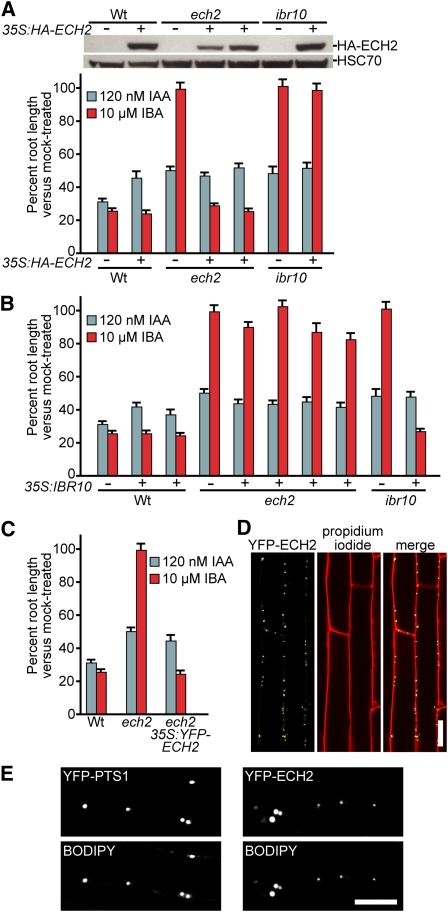
Figure 3.
ech2 Is Rescued by ECH2 but Not IBR10.
(A) Overexpression of HA-tagged ECH2 restores IBA sensitivity to ech2-1 but not ibr10-1. Top: Immunoblot analysis of (left to right) 5-d-old light-grown wild type (Wt), wild type carrying 35S:HA-ECH2, ech2-1, two independent ech2-1 lines carrying 35S:HA-ECH2, ibr10-1, and ibr10-1 carrying 35S:HA-ECH2. Anti-HA and anti-HSC70 antibodies were used to detect HA-ECH2 and HSC70 (loading control), respectively. Bottom: Mean normalized root lengths (+se; n ≥ 11) of 8-d-old light-grown lines shown in the immunoblot above the graph.
(B) Overexpression of IBR10 restores IBA sensitivity to ibr10-1 but not ech2-1. Mean normalized root lengths (+se; n ≥ 9) of 8-d-old light-grown wild type, two independent wild-type lines carrying 35S:IBR10, ech2-1, four independent ech2-1 lines carrying 35S:IBR10, ibr10-1, and ibr10-1 carrying 35S:IBR10.
(C) YFP-tagged ECH2 rescues ech2-1. Mean normalized root lengths (+se; n ≥ 11) of 8-d-old light-grown wild type, ech2-1, and ech2-1 carrying 35S:YFP-ECH2.
(D) YFP-ECH2 localizes to punctate structures in Arabidopsis cells. Confocal images of root epidermal cells from 4-d-old ech2-1 expressing YFP-ECH2 counterstained with propidium iodide to visualize cell walls. Bar = 20 μm.
(E) YFP-ECH2 localizes to peroxisomes in Arabidopsis cells. Confocal images of root epidermal cells from 4-d-old wild type expressing a peroxisomally targeted YFP derivative (YFP-PTS1; left panels) (px-yk; Nelson et al., 2007) and ech2-1 expressing YFP-ECH2 (right panels). The top panel of each pair shows the fusion protein fluorescence and the bottom panel of each pair shows fluorescence from 8-(4-nitrophenyl)-BODIPY, which allows visualization of peroxisomes (Landrum et al., 2010). Bar = 10 μm.

