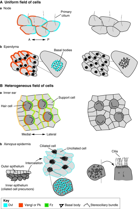
Fig. 6.
Organisation of vertebrate planar polarity in uniform and heterogeneous fields of cells. (A) Planar polarity in uniform fields of cells. In all panels, protein distributions are schematised on the left where known: Dvl (Dishevelled; blue), Vangl (Vang-like) or Pk (Prickle; orange), Fz (Frizzled; green). Surface views shown except where noted. The arrangements of polarised cells as seen in planar polarity mutants are illustrated on the right. In (a) the vertebrate node and (b) the ependymal lining of the brain, identically polarised cells point in one direction, as revealed by the asymmetric position of primary cilia in the node and patches of cilia in the ependyma (translational polarity). Planar polarity also exists at the subcellular level – i.e. rotational polarity – as revealed by the alignment of basal bodies (arrowheads) in individual ependymal cells (b). Cilia remain in the centre and/or fail to align with each other when planar polarity is not properly established. (B) In heterogeneous fields of cells, such as (a) the mouse inner ear and (b) Xenopus epidermis, polarised cells interdigitate with cells that show no outward polarisation. In the inner ear, core proteins are asymmetrically distributed in both hair cells (dark grey) and support cells (light grey); protein distributions are represented in lighter shades in support cells. In polarity mutants, hair cells are not uniformly oriented. In the epidermis, ciliated cells are born in the inner epithelium and then intercalate with unciliated cells in the outer epithelium (shown in cross-section), creating a mosaic of polarised and unpolarised cells in the mature epithelium (shown in surface view). Dvl and Vangl proteins may localise to basal bodies, which normally all point in the same direction. In planar polarity mutants, ciliary orientation is disrupted; defects in ciliogenesis and the apical localisation of the basal bodies have also been reported, as illustrated for one cell shown in cross-section on the right. A, anterior; P, posterior.

