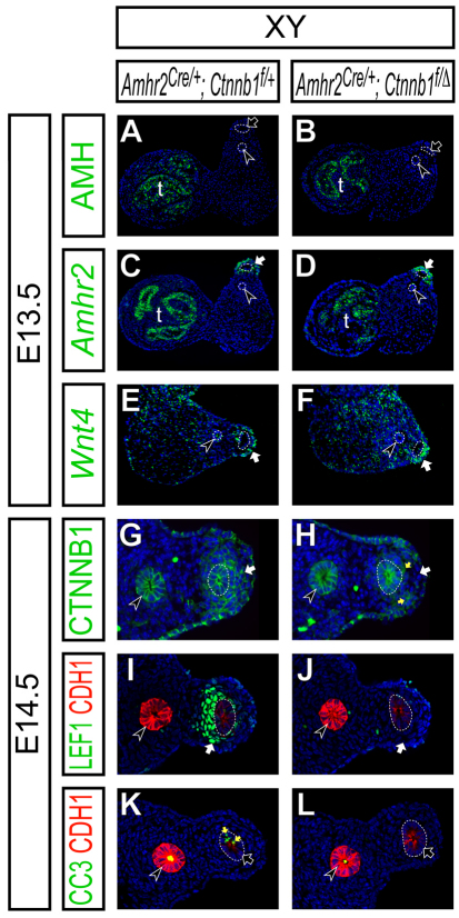
Fig. 7.
AMH signaling is activated normally in the mouse Müllerian duct mesenchyme in the absence of β-catenin activity. (A-L) Molecular marker analysis in the reproductive organs at E13.5 (A-F) and reproductive tracts at E14.5 (G-L) in Amhr2Cre/+; Ctnnb1flox/+ (A,C,E,G,I,K) and Amhr2Cre/+; Ctnnb1flox/del (B,D,F,H,J,L) XY male embryos. (A,B) Epifluorescence immunostaining of AMH (green) with DAPI staining (blue). (C-F) Radioactive section in situ hybridization for Amhr2 (green in C,D) and Wnt4 (green in E,F) with Hoechst staining (blue). (G-L) Confocal immunofluorescence. (G,H) β-catenin (Ctnnb1, green) with DAPI (blue) staining. (I,J) LEF1 (green), E-cadherin (CDH1, red) with Hoechst (blue) staining. (K,L) Cleaved caspase-3 (CC3, green), E-cadherin (CDH1, red) with Hoechst (blue) staining. E-cadherin is highly expressed in the Wolffian duct epithelium (black arrowheads) and weakly in the Müllerian duct epithelium (dotted line) in I-L. White arrows, black arrows, black arrowheads and yellow arrows indicate the Müllerian duct mesenchyme, Müllerian duct epithelium, Wolffian duct epithelium and cleaved caspase-3 positive apoptotic cells, respectively. t, testis.

