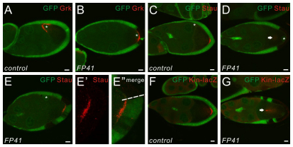
Fig. 1.
Oocyte polarity is disrupted in the presence of FP41 mutant PFCs. (A,B) Drosophila stage 9 egg chambers stained for Grk (red). The oocyte nucleus is marked by the white asterisks. Wild-type follicle cells express GFP (green); FP41 mutant cells are marked by the absence of GFP. In wild-type egg chambers, the oocyte nucleus and Grk protein are localized to the dorsal anterior corner of the oocyte at stage 9 (A). With the presence of FP41 mutant clones at the posterior, the oocyte nucleus and Grk are mislocalized at the posterior of the oocyte (B). Note that in this egg chamber the polar cells are wild type, as marked by GFP expression. (C-E″) Stage 9 egg chambers stained for Stau (red). The oocyte nucleus is marked by the white asterisks. In wild-type egg chambers, Stau protein is localized in a crescent at the posterior pole of the oocyte at stage 9 (C). When all the PFCs are mutant for FP41, Stau (arrow) is dispersed in the middle of the oocyte (D). When a portion of the PFCs are mutant for FP41, Stau is localized as a crescent in the region of the oocyte precisely underneath the wild-type PFCs (E). E′ and E″ are higher magnifications of E with a focus on the PFCs. The boundary between the mutant and the wild-type clones is marked by a dashed line. (F,G) Stage 9 egg chambers expressing the microtubule (MT) plus-end marker kin-lacZ and stained for β-galactosidase (red). In wild-type egg chambers, β-galactosidase is localized in a crescent at the posterior pole of the oocyte at stage 9 (F). With the PFCs mutant for FP41, β-galactosidase (arrow) is mislocalized in the middle of the oocyte, showing that the oocyte MT cytoskeleton is mispolarized (G). Scale bars: 10 μm. Egg chambers are oriented with the posterior side to the right.

