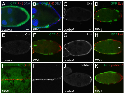
Fig. 2.
The FP41 mutation disrupts cell differentiation and Notch signaling in PFCs. (A-H) Stage 9 egg chambers stained for PH3 (A,B), Eya (C,D), Cut (E,F) or Hnt (G,H). FP41 mutant follicle cell clones are marked by the absence of GFP (green). (A,B) In wild-type egg chambers (A), the follicle cells stop dividing and switch from mitotic cycles to endocycles upon activation of Notch signaling. No mitotic cells labeled by PH3 (red) can be observed after stage 6. PH3-postive cells were still detected in FP41 mutant PFCs at stage 9 (B), indicating a failure of these cells to switch from mitosis into endocycle. Cell nuclei are stained with Hoechst (blue). (C,D) Eya expression is restricted to the anterior follicle cells at stage 9 in wild-type egg chambers (C). Eya (red) is still expressed in FP41 mutant PFCs (D), showing that cell differentiation is affected. (E,F) In wild-type egg chambers (E), Cut is normally downregulated in follicle cells at stage 7 by Notch signaling and only expressed in polar cells afterwards. In egg chambers containing FP41 mutant follicle cells at stage 9 (F), Cut (red) is still expressed but is restricted to the posterior mutant clones. (G,H) In wild type (G), Hnt expression is induced in all the follicle cells when Notch signaling is activated at stage 7. Hnt (red) fails to be upregulated exclusively in FP41 mutant PFCs (H, arrow) whereas other mutant follicle cells express Hnt normally. (I,I′) A wing disc containing FP41 mutant clones marked by the absence of GFP (green) and stained for Cut (red). The expression pattern of Cut is not affected in FP41 mutant cells in wing discs. (J,K) Stage 9 egg chambers expressing the posterior cell fate marker pnt-lacZ, and stained for β-galactosidase (red). Upon activation of the Egfr pathway, β-galactosidase staining reveals that pnt-lacZ is expressed in the induced PFCs in wild-type controls (J). FP41 mutant PFCs, marked by the absence of GFP (green), are able to express pnt-lacZ and respond to Egfr signaling (K). Scale bars: 10 μm. Egg chambers are oriented with the posterior side to the right.

