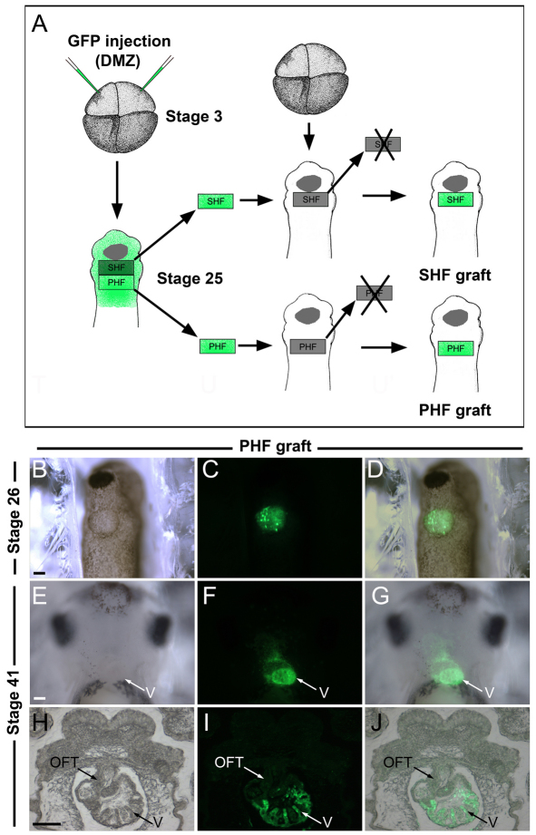
Fig. 4.
The PHF contributes to the ventricle. (A) Experimental design to analyze the relative contribution of the PHF and SHF to the developing heart. Four-cell stage Xenopus embryos were injected in the dorsal marginal zone (DMZ) with mRNA encoding GFP. At stage 25, the GFP-labeled PHF or SHF was transplanted onto the equivalent region of an unlabeled host embryo (PHF graft or SHF graft). (B-D) Ventral view (anterior to top) of a host embryo at stage 26 shows the position of GFP-labeled PHF graft. (E-G) At stage 41, the GFP-labeled PHF-derived cells are detected in the developing heart. Ventral view, anterior to top. (H-J) In a transverse section, the GFP-labeled PHF-derived cells are largely confined to the ventricle. OFT, outflow tract; V, ventricle. Scale bars: 100 μm.

