Fig. 7.
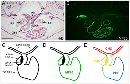
The relative contribution of the cardiac NC and SHF to the Xenopus cardiovascular system. (A) Hematoxylin and Eosin (H/E) staining of a transverse section through the heart of a stage 50 embryo. AA, aortic arch arteries; AS, aortic sac; LA, left atrium; OFT, outflow tract; RA, right atrium; SS, spiral septum; V, ventricle. Scale bar: 100 μm. (B) An adjacent section stained with MF20 antibody highlights the very sharp boundary between the muscle cells in the myocardium of the outflow tract and the aortic sac. (C) The Xenopus developing heart. (D) The muscle marker MF20 is expressed in muscle cells of the myocardium, including outflow tract and ventricle, but is excluded from the spiral septum. (E) The structures derived from the cardiac NC (CNC, red), the SHF (yellow) and the PHF (blue) are indicated based on our findings. PHF and SHF overlapping regions that result from the incomplete segregation of the two heart fields at the time of transplantation are in green.

