Fig. 2.
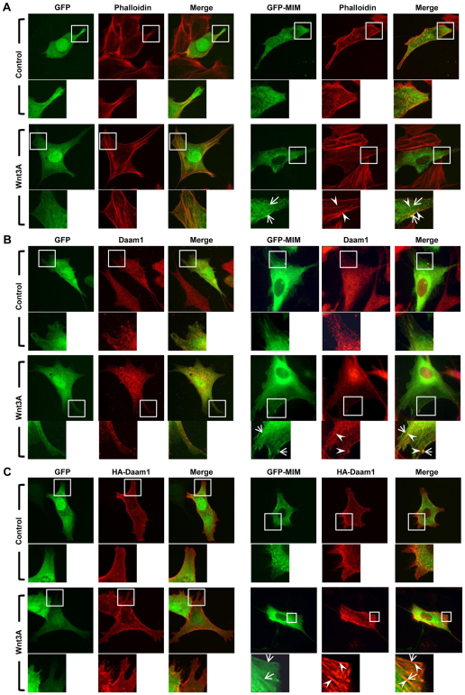
Wnt stimulation induces co-localization of Daam1 and MIM. (A) GFP-MIM, but not GFP, is localized to actin fibers and the plasma membrane upon Wnt3a stimulation. (B,C) Endogenous Daam1 (B) or HA-Daam1 (C) (red) co-localizes with GFP-MIM, but not GFP, to structures resembling actin fibers (arrowheads) and at the plasma membrane (arrows) upon Wnt3a stimulation. Boxes indicate the regions magnified beneath. Mouse NIH3T3 cells were cultured in the presence of 10% serum and Wnt3a stimulation was for 3 hours.

