Fig. 3.
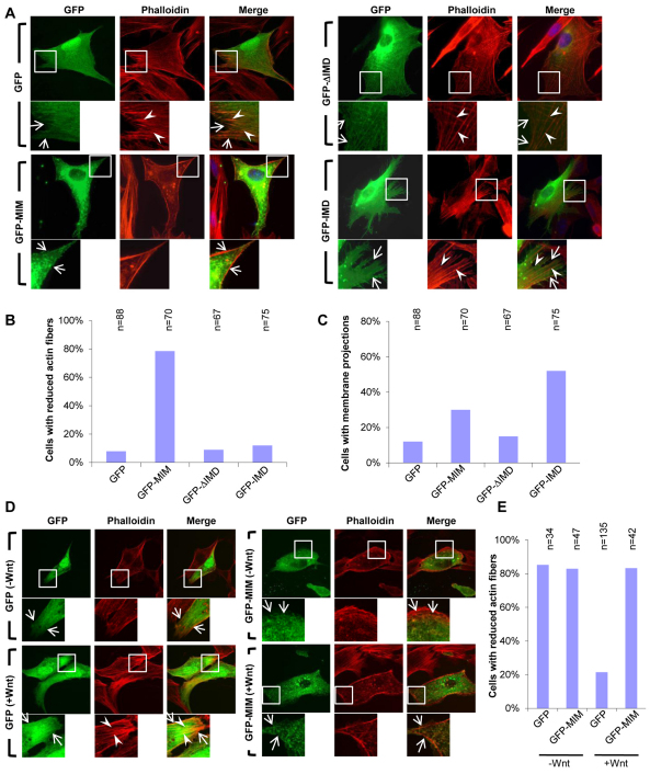
MIM regulates cellular actin fiber integrity and the formation of membrane protrusions. (A) GFP-MIM, especially GFP-ΔIMD, strongly co-localizes with Phalloidin-stained actin fibers (red). However, GFP-IMD only partially localized to, or decorated, actin fibers (arrowheads), but induced numerous membranous protrusions (arrows). Cells transfected with GFP were used as controls and transfected cells were cultured in the presence of 10% serum. (B) Quantification of the effects of GFP-MIM, GFP-ΔIMD and GFP-IMD on the amount of actin fibers within transfected cells. (C) Quantification of the ability of GFP-MIM, GFP-ΔIMD and GFP-IMD to induce membrane projections within transfected cells. (D) Transfection of GFP-MIM but not GFP inhibits Wnt3a-mediated actin fiber induction. cDNAs were transfected into serum-starved NIH3T3 cells for 24 hours with 3 hours Wnt3a stimulation. (E) Quantification of the results of D. The number of cells analyzed in B, C and E are shown above each bar.

