Fig. 7.
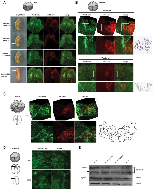
Depletion of MIM affects anterior neural fold elevation, hinge point formation and apical constriction. (A) MIM depletion inhibits neural fold elevation and closure only in the anterior of the Xenopus embryo. Arrows indicate where sectioning was performed. Transverse sections of a stage 22 embryo show the actin cytoskeleton stained with Phalloidin (green), and mCherry (red) labels cells injected with control MO or MIM MO. Neural fold elevation is only affected in the anterior of the embryo and not in the medial or posterior regions. (B) MIM is required for hinge point formation. In control MO-injected embryos, the anterior neural plate bends at two paired lateral hinge points (not shown). In embryos injected unilaterally with MIM MO, no hinge point forms on the injected (right-hand) side. Actin staining confirms the absence of a hinge point on the MIM MO-injected side (boxed). Drawings illustrate the cell shape on the control MO-injected and MIM MO-injected sides. Asterisks indicate injected side. (C) Strong actin staining is shown on the uninjected side, but no actin accumulation is found on the MIM MO-injected side (boxed). Drawing illustrates the cell shape on the control MO-injected and MIM MO-injected sides. Uninjected cells adopt elongated, polarized morphologies, whereas MIM MO-injected cells are short, round and disoriented. (D) Examination of cells within the anterior neural fold reveals that depletion of MIM inhibits actin fibers, resulting in disrupted morphology. (E) Western blotting shows that MIM preferentially interacts with endogenous Daam1 in the anterior of the embryo. Myc-MIM RNA was injected dorsally and whole embryo or sectioned embryo lysates were isolated at stage 16 and stage 18.

