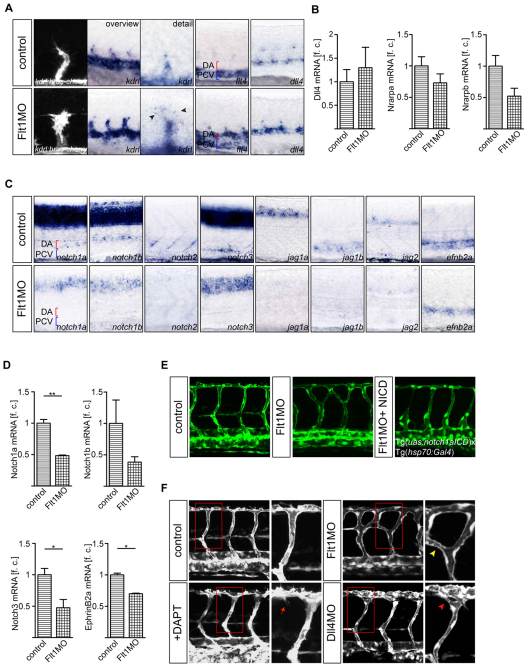
Fig. 4.
Tip and stalk cell marker expression and Notch signaling in flt1 morphants. (A,B) Expression of kdrl, flt4 and dll4 in control and flt1 morphants. Expression of the tip cell marker kdrl in segmental sprouts in Tg(kdrl:hras-mcherry)s896 transgenic zebrafish embryos (A, left) and kdrl in toto in situ hybridization (A, overview and detail). Note the ectopic expression of flt4 in the dorsal aorta of flt1 morphants (A, fourth panel). Expression of the stalk cell markers nrarpa and nrarpb is reduced, but expression of dll4 is maintained in morphants (A, fifth panel; B). (C,D) In situ hybridization and Taqman analysis of the trunk region show reduced expression of the receptors notch1a, notch1b, notch2 and notch3 and of the Notch ligands jag1a, jag1b and jag2 in flt1 morphants. Reduced expression of efnb2a suggests loss of Notch signaling in flt1 morphants. (E) Conditional overexpression of notch1a intracellular cleaved domain (NICD) in flt1 morphants (right) rescues segmental arterial branching defects. (F) Segmental branching pattern in dll4 morphants (red arrowhead) or in embryos treated with the Notch γ-secretase inhibitor DAPT (red arrow) did not phenocopy the branching pattern of flt1 morphants (yellow arrowhead). The boxed regions are shown at higher magnification in the right-hand panels. DA, dorsal aorta; PCV, posterior cardinal vein. *, P<0.05; **, P<0.01; Student's t-test. Error bars indicate s.e.m. f.c., fold change relative to age-matched control embryos.

