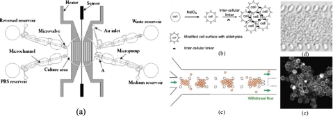
Figure 3.
(a) Schematic illustration of a 2D, automatic, cell-culture system including a cell-culture area, four micropumps, four microcheck valves, microchannels, reservoirs, two heaters, and a microtemperature sensor (Ref. 14). [(b)–(e)] A 3D, gel-free microfluidic device. (b) Cell surfaces modified by sodium periodate (NaIO4) have aldehyde groups which react with hydrazides on the intercellular linker to form multicellular aggregates. (c) Cells are suspended in a cell-culture medium with a dissolved intercellular linker and are seeded into the microfluidic channel with an exit flow at the outlet. (d) Transmission image of the cellular construct in a gel-free 3D-mFCCS after seeding. (e) Confocal microscopic image of cells aggregated with fluorescent intercellular linkers. Additional details regarding the fabrication of devices and experimental procedures can be found in Ref. 74. [Reprinted with permission from S. M. Ong et al., Biomaterials 29, 3237 (2008). © 2008, Elsevier.]

