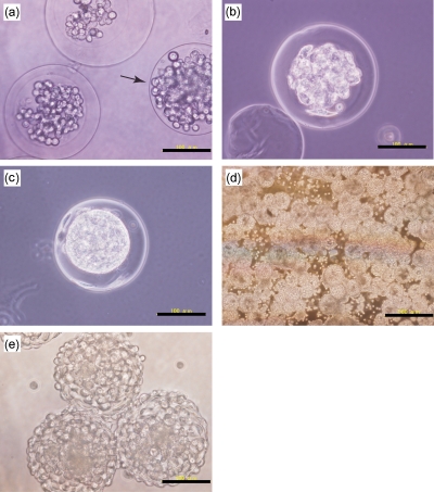
Figure 4.
Microphotographs of rat adipose-derived cells-enclosing ECGelatin-HPh microparticles (a) just after encapsulation of unmodified gelatin microparticles, after (b) 1 and (c) 4 days of incubation, and 1 day after seeding L929 cells on HepG2 cell-enclosing ECGelatin-HPh microcapsules at (d) low magnification and (e) high magnification. The arrow in panel (a) indicates the cell-enclosing unmodified gelatin microparticles without ECGelatin-HPh gel membrane. Bars indicate the scale: either 100 or 500 μm.

