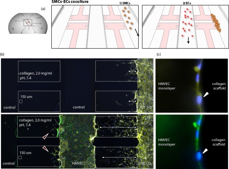
Figure 2.
Microfluidic device for culturing mural cells and endothelial cells. (a) Coculture model including mural cells and EC (HMVEC). (b) SMCs are cultured in the right channel and ECs are cultured in the center channel. The presence of ECs increased 3D invasion of SMCs into the collagen type I ECM, while ECs become stabilized by the presence of SMC (Ref. 21). (c) Adhered SMCs on the HMVEC monolayer, shown by a white arrowhead. Nucleus (DAPI; blue) and actin filaments (rhodamine phalloidin; yellow). Green antibody staining indicates HMVECs.

