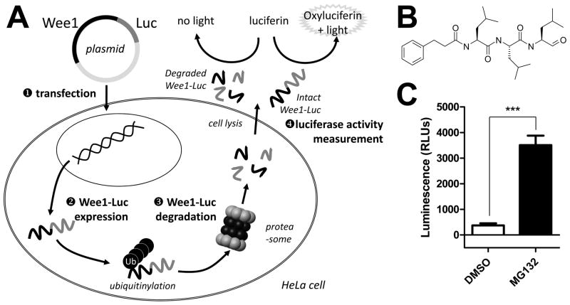
FIG. 1.
(A) HeLa cells were transfected with a plasmid allowing the expression of a chimeric Wee1 protein fused to the firefly luciferase at its C-terminal end. Upon ubiquitinylation, the Wee1-Luc fusion protein is subjected to degradation through the ubiquitin-proteasome pathway. Wee1-Luc protein levels were determined by measuring the conversion of the luciferase substrate D-luciferin into light-emitting oxiluciferin after cell lysis. A compound able to prevent Wee1 degradation is detected by an increase in measured luminescence. Ub: ubiquitin; Luc: luciferase; Wee1-Luc: K328M-Wee1-Luc. (B) Structure of the reference compound MG132. (C) Raw luminescence counts measured on the ViewLux (PerkinElmer) after HeLa cells have been transfected with the K328M-Wee1-Luc expressing plasmid and treated with DMSO or 20 μM MG132. Bars represent the average ± SD (n=4). *** indicates a P value greater than 0.0001 by a paired t test. The actual P value was 0.0007 and the calculated Z′ was 0.56.

