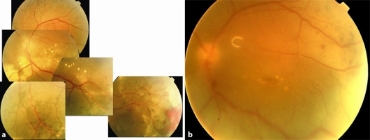
Fig. 2.
a Fundus photograph showing pigmentary changes with abnormal vasculature and lipid exudation involving the macula. Note the hemorrhage in the inferotemporal quadrant. Visual acuity was hand motions. b Postoperative fundus photograph of the same patient. The retina was attached and there was no lipid exudation. The visual acuity was 20/400.

