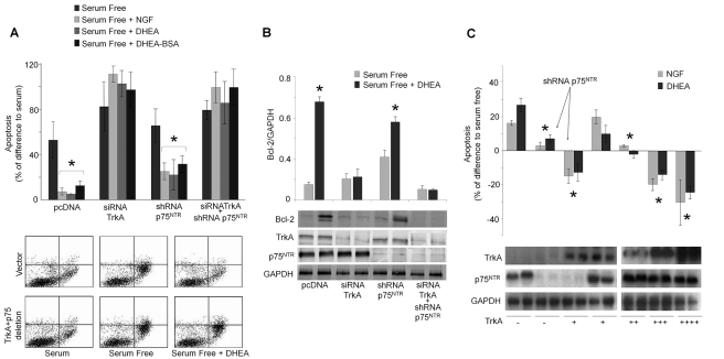
Figure 1. RNA interference against NGF receptors reverses the anti-apoptotic effect of DHEA.
PC12 or PC12nnr5 cells were transfected with si/shRNAs of TrkA and/or p75NTR (A and B) and/or expressing vectors of TrkA (c). Twenty-four hours later the medium was replaced either with complete medium (serum supplemented) or serum free medium, in the absence or the presence of DHEA, DHEA-BSA (100 nM), or NGF (100 ng/ml). Apoptosis was quantified 24 h later by FACS using Annexin V-FITC and PI. (A) Upper panel: levels of apoptosis expressed as % of difference from serum supplemented cells [* p<0.01 versus control (serum conditions), n = 8]. Lower panel: representative FACS analysis of Annexin V-FITC and PI staining. (B) Levels of Bcl-2 protein in serum deprived PC12 cells with or without DHEA treatment. Cellular extracts containing total proteins were collected and levels of Bcl-2 protein were measured by Western blot, and normalized per GAPDH protein content. Upper panel: mean ± SE of Bcl-2 levels, normalized against GAPDH (* p<0.01 versus control, n = 4), lower panel: representative Western blots of Bcl-2, TrkA, p75NTR, and GAPDH proteins. (C) Upper panel: levels of apoptosis in PC12nnr5 cells expressed as % of difference from serum deprivation condition. (* p<0.01 versus control-naive cells, n = 4). Lower panel: Western blots of TrkA, p75NTR, and GAPDH proteins for each condition.

