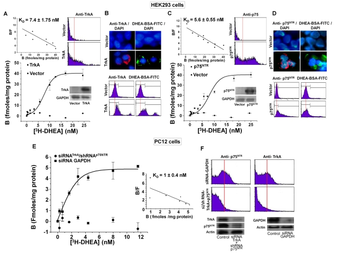
Figure 3. DHEA binds with high affinity to HEK293TrkA and HEK293p75NTR cell membranes.
(A, C) [3H]-DHEA saturation binding assays and Scatchard blots in HEK293 cells, transfected with the plasmid cDNAs of TrkA and p75NTR receptors. Fifty µl of cell membrane suspension in triplicate were incubated for 30 min at 25°C with 1–30 nM [3H]-DHEA in the presence or absence of 500-fold molar excess of DHEA. Western blot inserts show the efficacy of transfection (KD represents the mean ± SE of 3 experiments) (E) [3H]-DHEA saturation binding assays in PC12 cells, transfected with the si/shRNAs against TrkA and p75NTR receptors or with the siRNA against GAPDH. The efficacy of transfection is shown in Western blot inserts and in FACS analysis (F), (KD represents the mean ± SE of three experiments). (B, D) Fluorescence localization of DHEA membrane binding on HEK293 cells transfected with the plasmid cDNAs of TrkA and p75NTR receptors. Transfectants were incubated with either the membrane impermeable DHEA-BSA-FITC conjugate (100 nM), BSA-FITC (100 nM), or with specific antibodies against TrkA and p75NTR proteins. Transfectants were analyzed under the confocal laser scanning microscope (B) or by FACS analysis (C). Blue staining depicts Hoechst nuclear staining.

