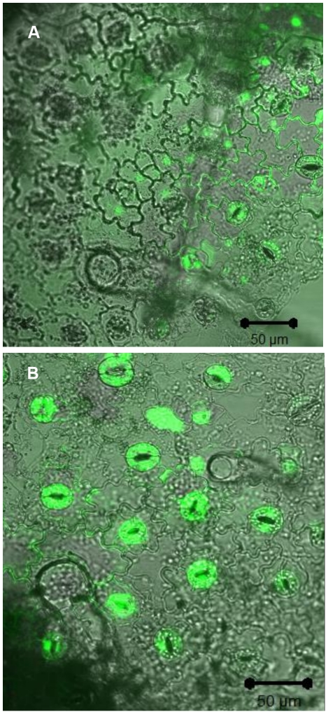
Figure 2. Laser scanning confocal imaging of the elicited- oxidative burst in the outmost epidermis cell layer of N. tabacum.
Epidermal tissues were loaded with H2DCF-DA, washed, and examined by laser scanning confocal microscopy. S. Typhimurium (A) and P. syringae (B) were added during time course of image acquisition. The images show green fluorescence mainly in the stomata guard cells. Image comparison evaluation demonstrates more than four times fluorescence intensity in image B. The laser setting, microscope filters and all other image parameters were identical in both images.

