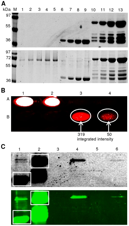
Figure 11. Protein-DNA interaction analysis based on IFP fusions.
(A) Fusion proteins ANAC042-TEV-IFP-6xHis (70 kDa) and IFP-6xHis (36 kDa) affinity-purified from E. coli. Two elution fractions (2×250 µL) containing the purified proteins were pooled and analysed after SDS-PAGE by in-gel detection (top) and Coomassie staining (bottom) (lanes 10–13: ANAC042-TEV-IFP-6xHis, 5, 10, 15, and 20 µL; lanes 6 - 9: IFP-6xHis, 2, 5, 8 and 10 µL). BSA served as standard to estimate protein amounts (lanes 1–5: 100/250/500/750/1000 ng). Equal amounts of both proteins (∼5 µg) were used for protein-DNA interaction analysis. M, molecular mass marker (kDa). (B) Biotinylated dsDNA was immobilized on streptavidin mutein particles and incubated with ANAC042-TEV-IFP-6xHis protein. After elution, fractions were scanned at 700 nm in the wells of a microtiter plate (strong infrared signal appears white in the digital image). A1: IFP-6xHis input. A2: ANAC042-TEV-IFP-6xHis input. A3: negative control; B-100%-DNA immobilized on streptavidin mutein particles + IFP-6xHis in the presence of non-biotinylated 7%-DNA. A4: negative control; B-7%-DNA immobilized on streptavidin mutein particles + IFP-6xHis, in the presence of non-biotinylated 100%-DNA. B1/2: empty wells. B3/4: experiments with B-100%-DNA and B-7%-DNA immobilized on streptavidin mutein beads + ANAC042-TEV-IFP-6xHis incubated in the presence of non-biotinylated 7%- and 100%-DNA, respectively. Areas of the infrared signals were marked (white circles) and integrated signal intensities were calculated (B3 = 319, and B4 = 50). (C) After infrared-scanning in microtiter plates (see B) samples were separated by SDS-PAGE and scanned at 700 nm (top) followed by western blot analysis (bottom). Lane 1: IFP-6xHis input (white square). Lane 2: ANAC042-TEV-IFP-6xHis input (white square). Lane 3: negative control with B-100%-DNA immobilized on streptavidin mutein particles + IFP-6xHis, in the presence of non-biotinylated 7%-DNA. Lane 4: experiment with B-100%-DNA immobilized on streptavidin mutein particles + ANAC042-TEV-IFP-6xHis, in the presence of non-biotinylated 7%-DNA. Lane 5: negative control with B-7%-DNA immobilized on streptavidin mutein particles + IFP-6xHis, in the presence of non-biotinylated 100%-DNA. Lane 6: experiment with B-7%-DNA immobilized on streptavidin mutein particles + ANAC042-TEV-IFP-6xHis, in the presence of non-biotinylated 100%-DNA.

