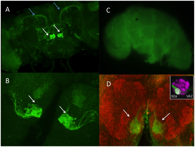
Figure 2. Psphinx-GFP line was stained with anti-GFP.
(A) antennal lobe (white arrowhead) and inner antennoglomerular tract (blue arrowhead); (B) zoomed-in image of the two glomeruli. (C) negative control for immunostaining. (D) Double-staining with anti-GFP to visualize sphinx expression in the glomerulus VA2 (green), and the synaptic marker mAb nc82 (red) to visualize the glomerular structure of the antennal lobe.

