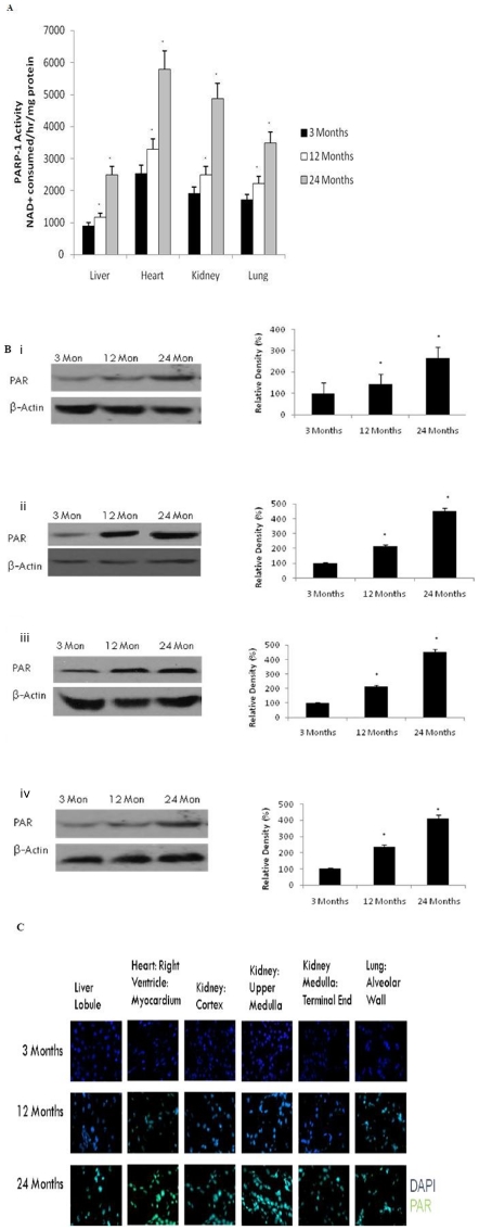
Figure 5. Increased Poly(ADP-ribose) activity in the brain with age.
(A) PARP activity was determined in aging tissue using a spectrophotometric assay. All values are means ± S.E from tissue obtained from eight different rats for each age group. Significance *p<0.01 compared to 3 month old rats. (B) Western blotting for poly(ADP-ribose) in (i) liver, (ii) heart, (iii) kidney, and (iv) lung with aging using anti-Poly(ADP-ribose) (10H) antibody. The blots shown are representative tracings of an experiment done eight times. Graphs are mean ± S.E from tissue obtained from eight different rats for each age group. Each bar of the quantification graph represents the corresponding band for each age group. Significance *p<0.01 compared to 3 month old rats. (C) Immunodetection for poly(ADP-ribose) in the liver, heart, kidney and lung from 3 month, 12 month and 24 month old rats. Poly(ADP-ribose) (green) and DAPI (blue). Higher immunoreactivity for poly(ADP-ribose) was observed in 12 and 24 month old rats compared to 3-month old rats.

