
Figure 3. Western blots against Hu.
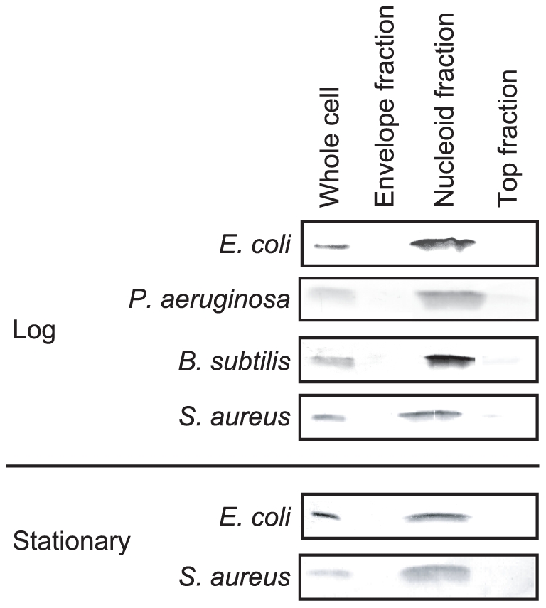
Five micrograms of proteins from the whole-cell lysates, the nucleoid fractions, the envelope fractions, and the top fractions were separated by SDS-PAGE, and Hu was detected by Western blotting. ‘Log’ and ‘Stationary’ represent the log phase and stationary phase, respectively.