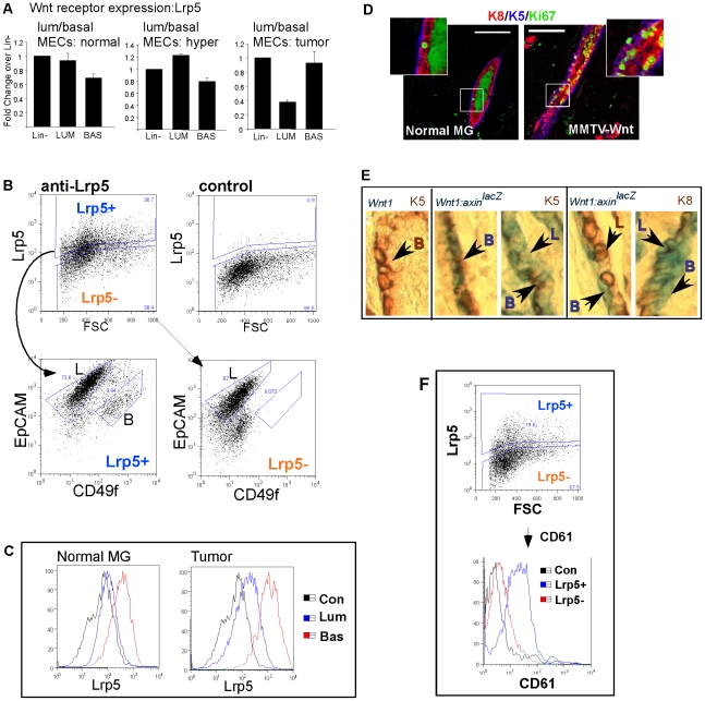
Figure 2. Lrp5 expression (acquired post-transcriptionally) correlates with the appearance of Axin2 in luminal cells.
(A) mRNA extracted from purified cell fractions was analyzed for relative expression of Lrp5 by quantitative RT-PCR analysis as described for Fig. 1. (Lrp6 mRNA expression is broadly similar to Lrp5, shown in Fig. S4) (B) Tumor cells were incubated with anti-Lrp5 antibody, and analyzed by flow cytometry, using an isotype-matched negative control to define Lrp5+ cells. These samples were also stained with EpCAM and CD49f antibodies, and the cell surface phenotypes combined to show the luminal (L) or basal (B) cell identity of Lrp5+ and Lrp5− cells. (C) Another example of this assay is shown in histogram form, to reveal the overall level of expression for Lrp5 in luminal and basal cells in normal and tumor cells. (D) Paraffin sections from normal and Wnt-induced mammary glands were stained to illustrate the relative rates of division (green, Ki67) of basal (blue, K5) and luminal (red, K8) cells. Note that the green color stain in the lumens is a common artifact associated with non-specific binding to sticky luminal proteins. Scale bar = 50 µm. (E) Axin2lacZ [MMTV-Wnt1] (and control Wnt1) glands were stained in whole mounts for the lacZ reporter, followed by embedding, sectioning and immunocytochemical assay of basal (K5+) and luminal (K8+) cells. The panel on the LHS shows there was no background when the Axin2lacZ reporter was not present. The samples were incubated in x-gal substrate (B, basal; L, luminal; as indicated). The pattern of staining of AxinlacZ was heterogeneous, in some areas, stain was basal-specific, in others, there was also light staining throughout the luminal population, in others, focalized clusters of luminal cells showed high expression. (F) Lrp5+ cells were assayed for their expression of CD61, because CD61+ cells have been shown to be enriched for TIC activity in this tumor model (Vaillant et al 2008). These markers showed high co-expression.

