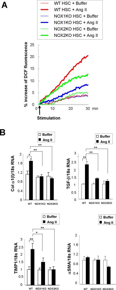
Fig. 7. Both NOX1 and NOX2 mediate ROS production and fibrogenic responses in HSCs.
(A) HSCs from WT, NOX1KO and NOX2KO mice were loaded with redox-sensitive dye CMH2DCFDA (10 μM) for 20 minutes. Cells were then washed twice and subsequently stimulated with angiotensin II (10−6 M). Fluorescent signals were quantified continuously for 30 minutes using a fluorometer. (B) mRNA expression of collagen α1(I), TGF-β, TIMP1, and α-SMA were measured in WT, NOX1KO, and NOX2KO mice 24 hours after angiotensin II (10−6 M) stimulation by qPCR. Data were from 2 independent experiments performed in triplicates. *P < 0.05, **P < 0.01.

