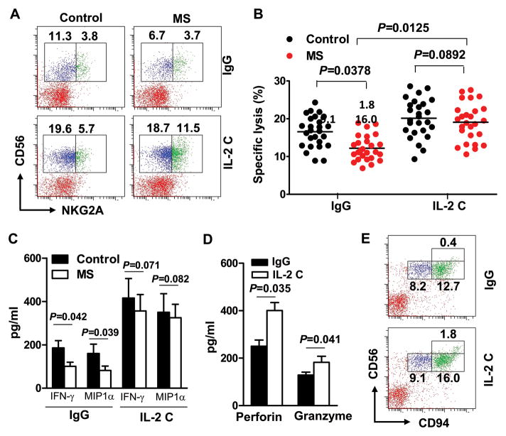
FIGURE 5. IL-2 complexes restore defective NK cells from MS patients.
Blood was drawn from relapsing remitting MS patients or healthy controls. Peripheral blood mononuclear cells (PBMC) were isolated and incubated with IgG or a combination of IL-2 (10 ng) and anti-IL-2 mAb (10 μg/ml) for 48–72 hours. (A) Frequency of CD56 and NKG2A double-positive cells on gated CD3− lymphocytes. Plots are representative of 26 MS patients and 26 controls. Each symbol represents one subject. P values, ANOVA test. (B) CD56dim cells were sorted after incubation with IgG or IL-2 complexes (IL-2 C) and incubated with 51Cr labeled K562 cells. Cytotoxicity toward K562 cells was measured by 51Cr release. (C) CD56bright cells were sorted after incubation with IgG or IL-2 C, production of IFN-γ and MIP-1α was quantified by ELISA. P<0.01 for comparisons between different cytokines in the control group and their corresponding IL-2 C treated groups. P values, ANOVA test. (D) Levels of perforin and granzyme B secreted by cultured human NK cells with IgG or IL-2 C were quantified by ELISA. The bar data represents results from two separate experiments (Mean and s.e.m.). P values, Student’s t-test. (E) Frequency of CD56 and CD94 double-positive cells on gated CD3−lymphocytes. Plots were representative of 6 MS patients.

