
Figure 1. Development of an orthotopic model with subsequent metastasis using human UCB cells.
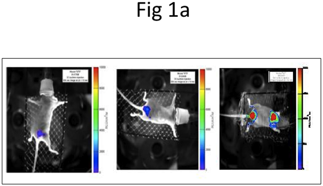
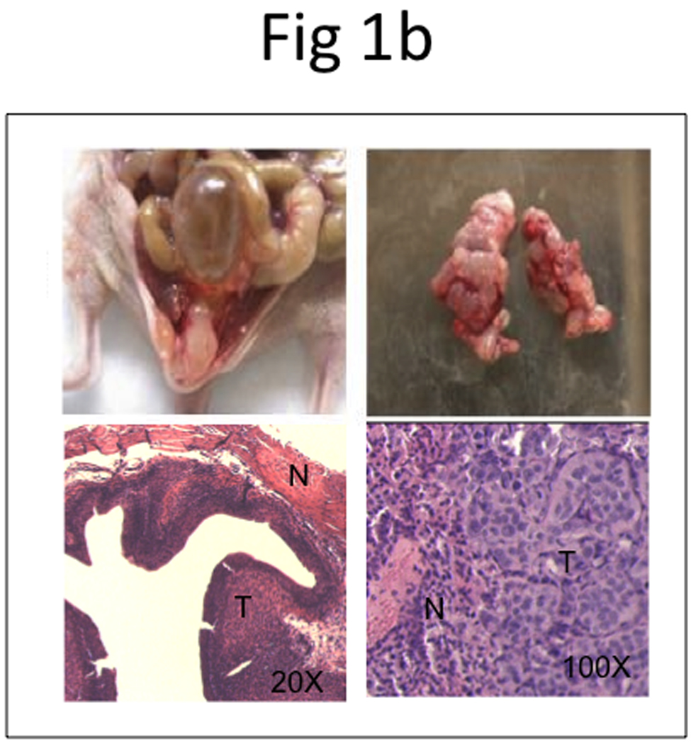
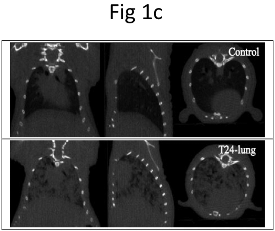
a, BLI imaging was carried out at Week 2 (left panel), 4 (middle panel), and 6 (right panel). b, Representative photos display primary tumor (top left panel) and histology of superficial tumor with an invasive phenotype (bottom left panel), lung metastases (top right panel) and histology of lung metastases (bottom right panel). N, normal; T, tumor. c, microCT of lung metastases was taken 42 days after bladder instillation.